Over the last three years or so I’ve gone through a ton of injuries. This might be because I’m getting older. Or that I’m taking more athletic risks. Here I want to discuss one such injury and document some of the damage and how it happened so that you, dear reader, can be smarter and learn from my mistakes. Or if you don’t learn, that you can see what these injuries look like and how to deal with them. This will be more of a show and tell and i will discuss my wrist ligament injury and how I localized and addressed it with and without a doctor’s help.
The Injury
So in October 2020, I injured myself. Generally, before this, I was doing about 40 pull-ups daily in a two sets – once in the morning and once at night. And things were going great. I was on top of the world and was getting a bit more bulky than my usual self.
But I made a mistake. I listened to a former amateur squash athlete. He had been close to becoming a championship player and well he was related. He suggested that I level up my pull-ups and change the position of my wrist so that I’m pulling up with my wrist area. So I thought, trying this once should be ok. And so, one fateful evening I tried this. And everything looked ok. Until the next day. In retrospect, I probably should not have listened to his advise considering his shortcomings from a point of reasonableness.
So what happened? I had injured the tendons or the ligaments or something in both my left and right wrists. At the time, however, I wasn’t sure what exactly had happened. I write this article a year and a half later. And well, they have gotten considerably, but not completely better. And I presently still experience tendonitis when playing the guitar and heavily using one of my wrists for DIY projects. If I ever happen to use my wrists with too heavy a load or if I sleep the wrong way it also encourages some level of discomfort. From the time of injury pull-ups were out of the question and so, as it turned out, were push-ups. Push-ups, were another thing I did 50 of everyday.
1. Likely Cause
The underlying cause for this injury was doing pull-ups wrong. What exactly was wrong with the pull-up you ask? Well, I’ve taken a couple of picture to demonstrate. Below are two pictures to explain.
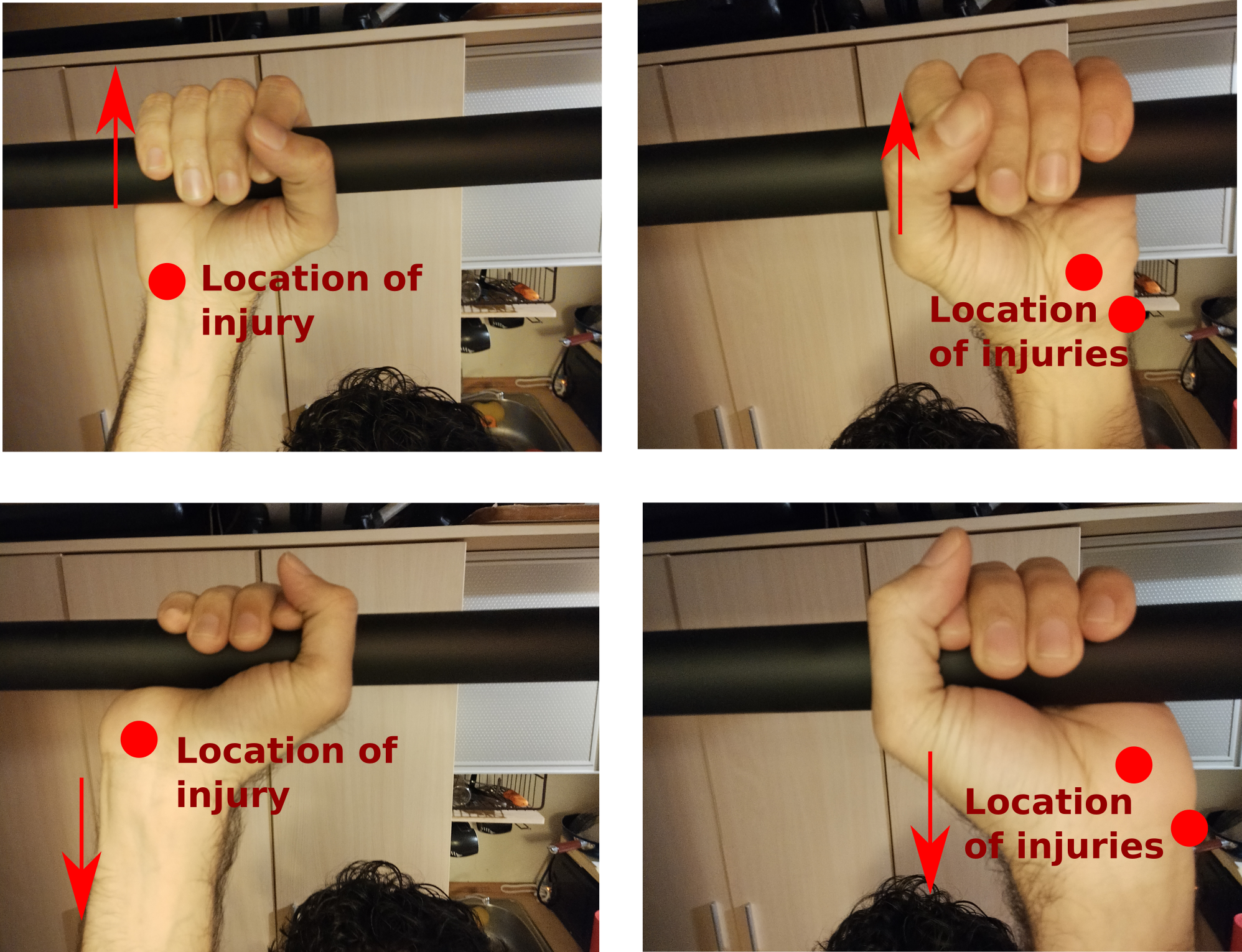
Essentially, what this set of pictures show is that I moved from the top row position while hanging off of my pull up bar to the bottom row position. I essentially did a mini-wrist pull up and brought my palms up to press against the pull up bar and then released. And a day or two later I began to notice injuries at the locations marked. These were difficult to localize in the beginning, but as time went on the pain became more localized.
2. Symptoms
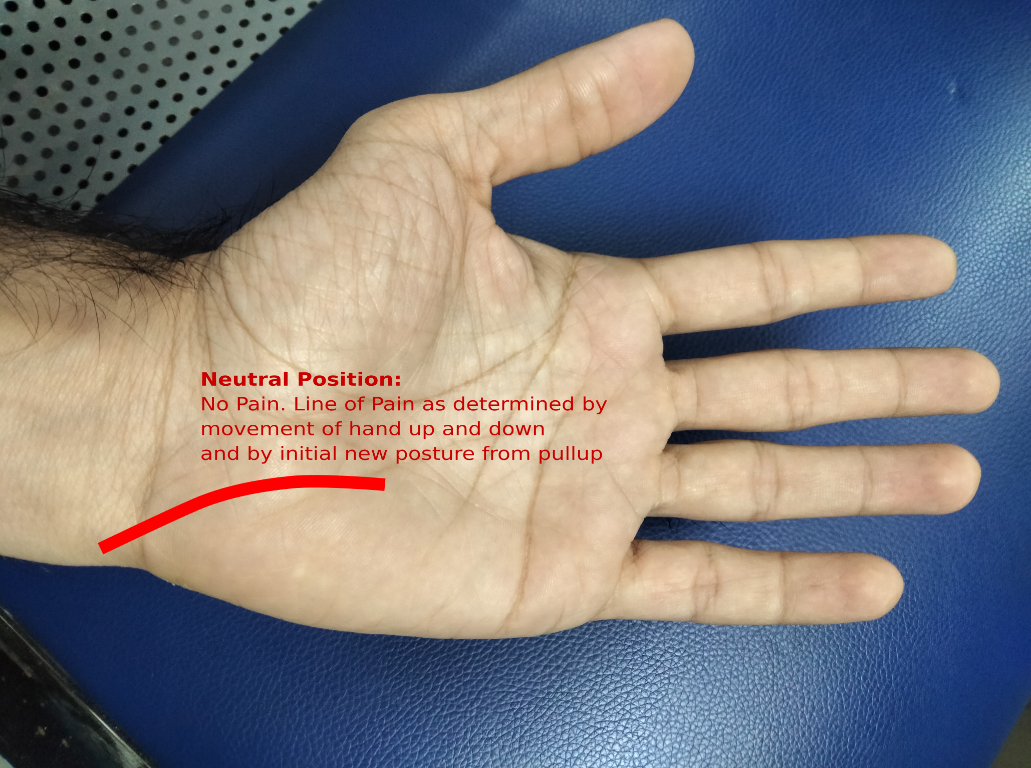
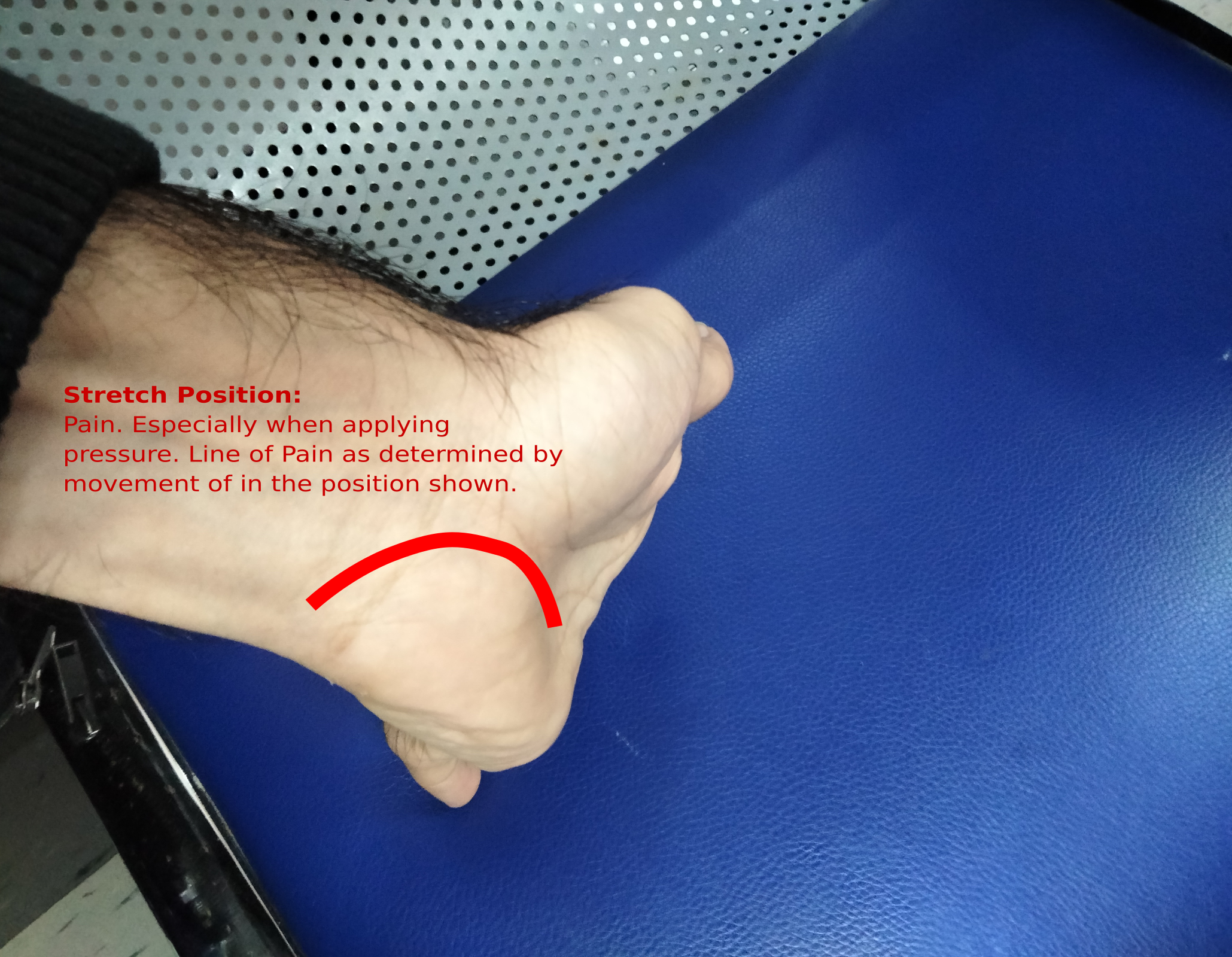
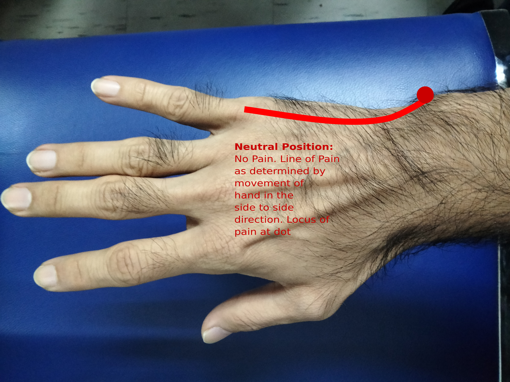
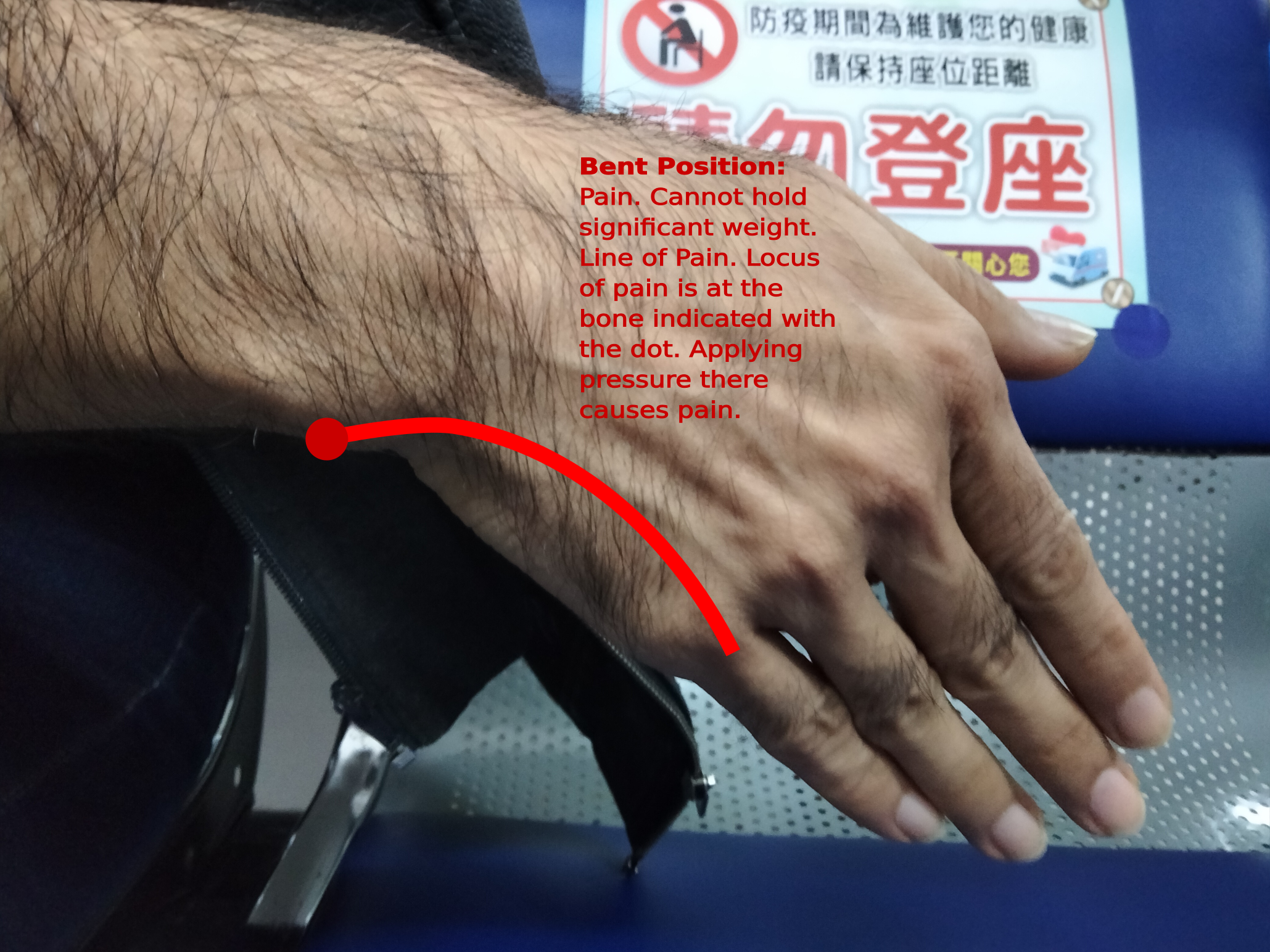
So the below set of images shows how I localized the injury locations. I took my hand and flexed it from a neutral position as shown for my left hand. In the neutral position, I experience no pain. However, as I moved to the stretched position it became painful.
The left hand healed faster and the right hand took longer. The left is mostly healed now with occasional aches after 1.5 years. However, the right hand has multiple injuries. One injury is similar to the left hand injury and that healed quickly. Significantly more quickly than the left hand.
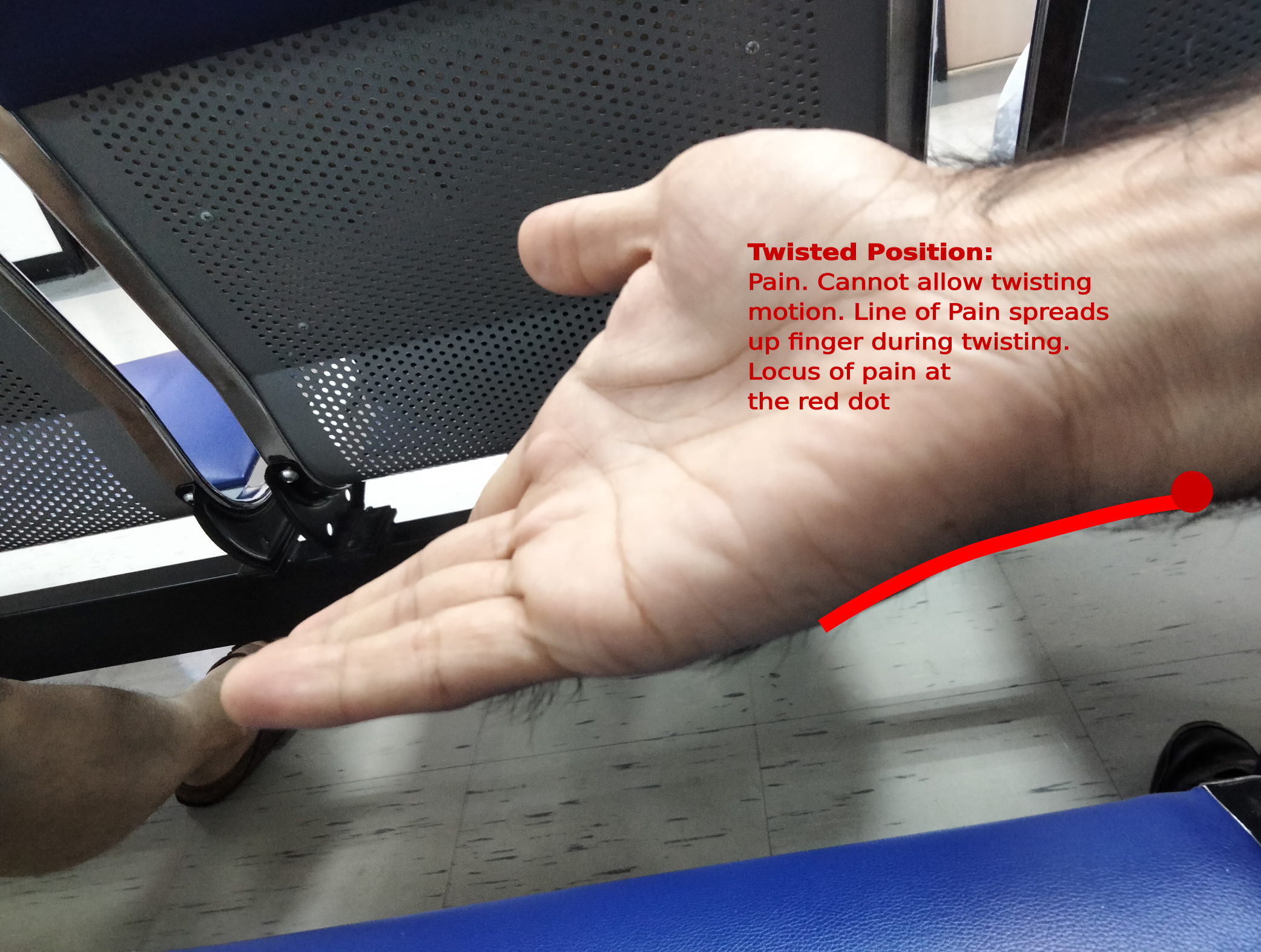
The additional injury in the right hand was and continues to be a problem to present day – though considerably more healed. I isolated this injury and localized it as shown in the pictures below moving from a no pain neutral position to a painful stretched and twisted position.
3. Identification of Specific Injury Location
To explain this, I’ll borrow some images from Elsevier’s Complete Anatomy software tool [1] for which I bought a professional license. I’ll be making use of these images in a non-commercial way for your education only and believe that they are fair use. Though I have some limited licensing rights as well.
The picture below shows an X-Ray done on one of my hands. The X-Ray isn’t a great tool for diagnosing soft tissue injury, I believe. However, according to the doctor, certain tears of tendons and ligaments can show up on X-Ray’s at the point of injury. I’m not sure how much I believe him just yet. The resulting X-Rays suggest though that the injury is not serious. Though considering my reduced mobility for such an extended period of time I would consider it somewhat serious. When an injury of this kind has such a long extended duration, I get the impression that it is sometimes called a chronic injury.
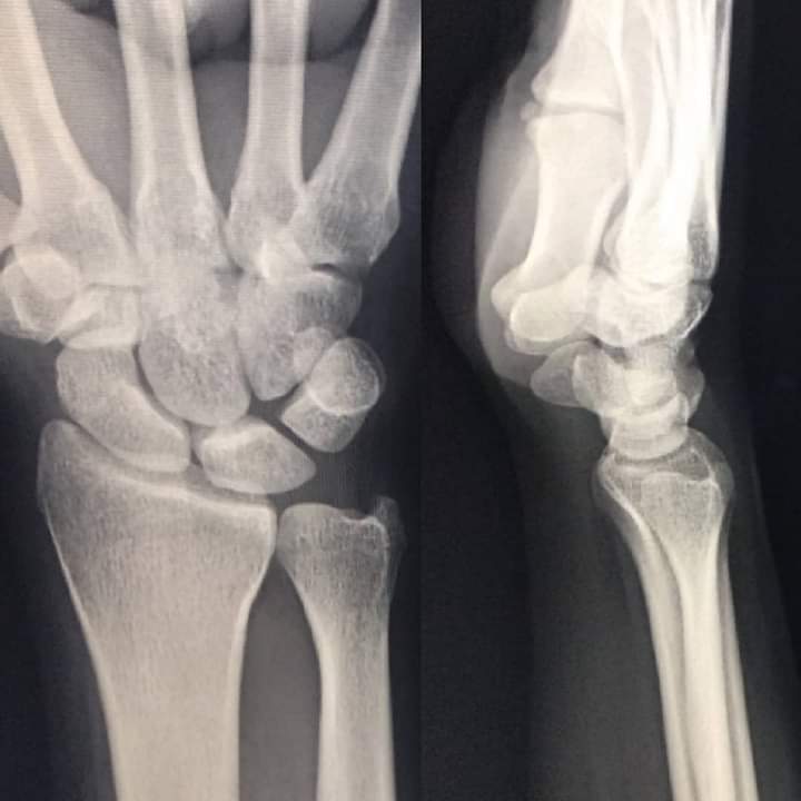
In my personal opinion, further analysis should have been done using some form of soft tissue imaging. Perhaps an MRI and ultrasound or similar. This soft tissue imaging could have helped clarify specific regions of the wrist that needed further attention. Though perhaps the reason for not examining further is a combination of not caring enough, having not enough time to pay attention to furthering the diagnosis or simply that further isolating the injury would not really help with the treatment.
I decided to make my own assessment because the doctor I saw was being arrogant and unwilling to carefully check the injury. I used Elsevier’s tool [1] and produced a comparison of an anatomical image of my wrist. with the regions of pain that I was experiencing.
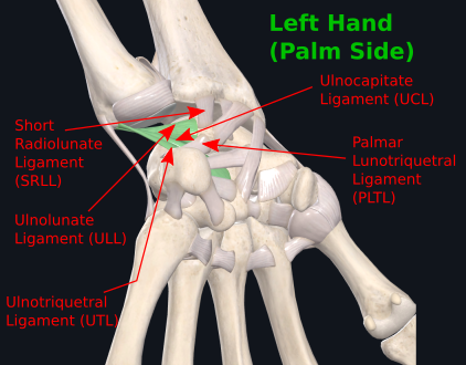
The image below is a closer look at my left wrist. A mirror image injury seems to have taken place on my right hand though it healed considerably in comparison to my left. I tried to locate the main sources of pain and identify possible ligaments that could be causing the pain. There were five main ligaments that might have been the issue.
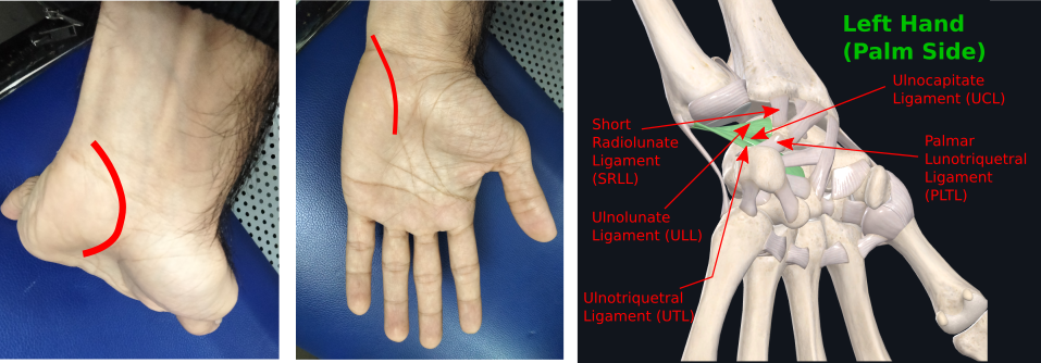
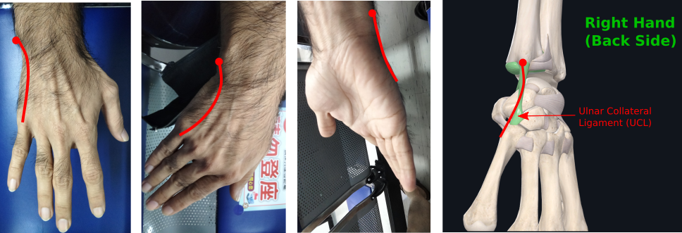
I repeated the same approach for my right hand. And the resulting image is shown below. However, in the image below I have only isolated the particular ligament that was causing pain as a result of what I suspect is an additional injury beyond that mirroring the left hand. This additional injury restricted my hand’s twisting motion.
4. Treatment
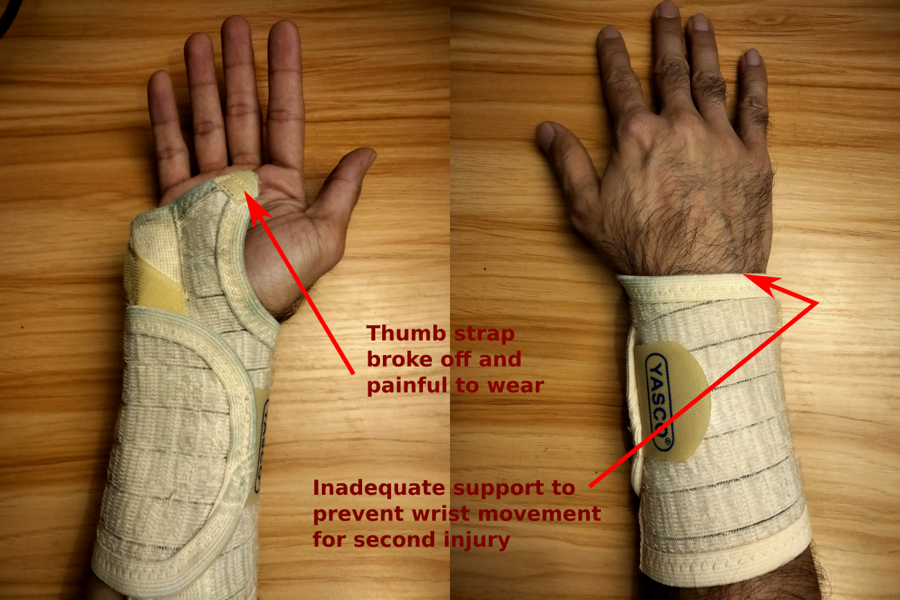
My first visit to the doctor was a month or two after the injury – this is when I started to get suspicious about why the wrists weren’t improving. During this month or so, I was already using heat and cold packs and a wrist brace that I had bought from a pharmacy.
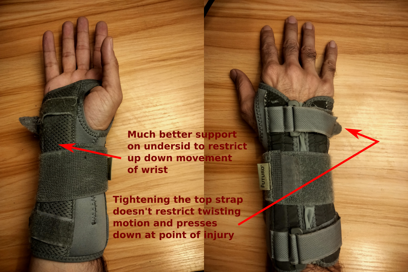
Upon visiting the doctor, he took the X-Ray, determined the problem was not serious and asked me to wear a more substantial wrist brace. A picture of my previous and new wrist brace is shown below. The real challenge with both these wrist braces was that they were not adequate in restricting motion of the wrists. Further, the first brace created pain around the base of my thumb that was unhelpful. And the second while significantly more stiff ended up causing more serious injury to the right hand by pressing against the outer bone (second injury of the right hand) as shown below. I also was not advised clearly on how long I should be wearing the wrist brace. I likely worsened the situation by wearing it to bed and for other extended periods of times causing the right wrist injury to get worse.
Subsequent to this I decided about 8 months later to go and see a rehabilitation specialist instead. She took considerably more time to assess the injury and on subsequent visits suggested a technique that I later looked up. This technique it seems is called prolotherapy and involves injecting a glucose solution directly at the point of injury into the ligament using a syringe. The picture below is an example of prolotherapy in action as taken from the online source of the Journal of Prolotherapy [2].
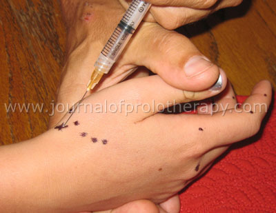
I had this done once, but I was told that I would need it several times. This was extremely painful and was done directly at the doctor’s desk. Searching the literature I realized that there was no clear evidence of its efficacy and decided not to repeat the procedure until I returned from Canada. The idea, as I understand it, behind the approach, is that it encourages inflammatory response at the location of the ligament injury and so allows higher blood flow to encourage healing. It is important not to take anti-inflammatory or painkilling drugs for this injury as it can limit the efficacy, apparently.
Finally the last of the interventions that the rehabilitation specialist recommended was cold laser therapy. Unfortunately at the time I was forced to cut short this treatment after one visit as a result of my trip to Canada. I haven’t been able to return to the physiotherapist since this time. I will publish some updates in a future articles to reflect my experiences here after a few visits. Bur for now here is an image of what cold laser therapy looks like in practice – not my image, taken from the web source [3].
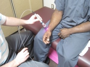
I believe the idea behind this form of treatment is that the laser penetrates through the top skin and muscle layers more easily at particular frequencies to provide heating directly to the ligament area or the light causes cells in the underlying layers to become stimulated to promote regeneration. But not sure quite yet.
5. References
- Elsevier Complete Anatomy Software. 2021. https://www.elsevier.com/solutions/complete-anatomy
- Rodney S. Van Pelt. Hand and Wrist Prolotherapy. November 2010. Teaching Techniques Volume 2, Issue 4. https://journalofprolotherapy.com/hand-and-wrist-prolotherapy/
- https://laserneuropathypaintreatment.com/conditions/carpal-tunnel/